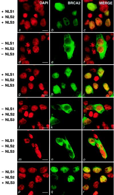
Figure 2.
Fluorescent confocal microscopy of cells expressing GFP-tagged full-length BRCA2 and deleted forms of BRCA2. 293T cells were transiently transfected with the following constructs: MG-B2GFP (a–c), MG-B2GFPΔ3263–3418 (d–f), MG-B2GFPΔ3270–3418 (g–i), MG-B2GFPΔ3263–70/Δ3381–85 (j–l), MG-B2GFPΔ3263–3385 (m–o) and MG-B2GFPΔ3263–3380 (p–r). (Left) DAPI was used to stain nuclei (red, a, d, g, j, m, p). (Center) Green fluorescence from the GFP fusions (green, b, e, h, k, n, q). (Right) The merged images are presented (c, f, i, l, o, r). To the left, the status of NLS1, NLS2, and NLS3 is indicated (+, present; −, absent). (Bar = 10 μm.)

