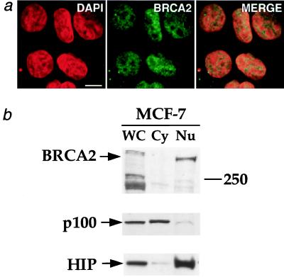
Figure 3.
Endogenous BRCA2 is nuclear. (a) Indirect immunofluorescence of human breast cancer cells (MCF-7) expressing full-length BRCA2. Cells were prepared as described in Materials and Methods, probed with anti-BRCA2A Ab (8) (BRCA2), stained with DAPI, and then imaged by confocal microscopy. The merged images are presented on the right. (Bar = 10 μm.) (b) Biochemical fractionation of MCF-7 cells. MCF-7 cells were separated into cytoplasmic (Cy) and nuclear (Nu) fractions. WC, whole-cell extract. Equal numbers of cells were used to prepare the whole-cell and fractionated lysates. (Top) Anti-BRCA2A Ab was used to probe for BRCA2. The 250-kDa protein marker band is marked on the right. To validate the fractionation procedure, separate blots of protein from the subcellular fractions were probed with antibodies against the cytoplasmic NF-κB2 p100 precursor protein (38) (Middle) and the nuclear HIP protein (39) (Bottom). These proteins fractionated as expected.

