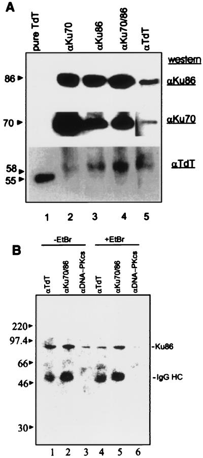
Figure 3.
(A) TdT–Ku interaction in Molt-4 lymphoblasts. Molt-4 lymphoid cell extracts (500 μg) were incubated with mAbs specific for Ku 70 (N3H10), Ku86 (111), Ku70/86 (162), or TdT (5100) as indicated above the lanes. Immunoprecipitated complexes were analyzed as described above. The blot was serially probed with rabbit anti-TdT (1:500 dilution; Bottom), mouse anti-Ku70 (1:1 dilution; Middle), or mouse anti-Ku86 antibodies (1:4,000 dilution; Top). The blot had to be exposed for a longer time to obtain a signal corresponding to Ku86. To prevent overexposure of other lanes, a separate exposure was obtained and included for lane 5. (B) Effect of EtBr on TdT–Ku interactions. Protein complexes were immunoprecipitated in the absence (lanes 1–3) or presence (lanes 4–6) of 100 μg/ml EtBr with anti-TdT, anti-Ku70/Ku86 heterodimer, and anti-DNA-PKcs mAbs (lanes 1 and 4, 2 and 5, 3 and 6, respectively) from Molt-4 cell extracts, resolved by SDS/PAGE, and analyzed by Western blotting by using anti-Ku86 as described in A.

