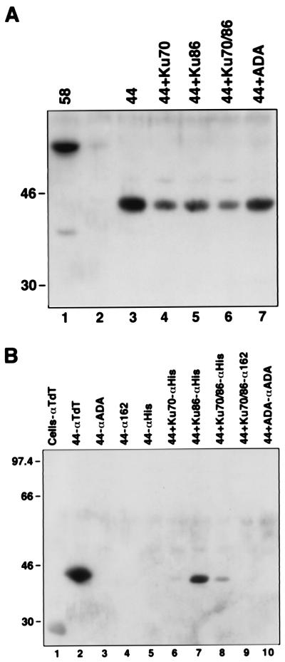
Figure 4.
(A) Expression of 44-kDa N-terminal truncation of TdT. Extracts from uninfected (lane 2) or baculovirus-infected Sf9 cells expressing 44-kDa TdT (lane 3), 44-kDa TdT with Ku70 (lane 4), 44-kDa TdT with Ku86 (lane 5), 44-kDa TdT with Ku70/86 heterodimer (lane 6), or 44-kDa TdT with ADA (lane 7) were analyzed by Western blotting by using rabbit polyclonal anti-TdT. (B) Coimmunoprecipitation of 44-kDa TdT with Ku86. Extracts from uninfected Sf9 cells (lane 1) or cells infected with baculovirus expressing 44-kDa TdT (lanes 2–5), 44-kDa TdT with Ku70 (lane 6), 44-kDa TdT with Ku86 (lane 7), 44-kDa TdT with Ku70/86 (lanes 8 and 9), or 44-kDa TdT with ADA (lane 10) were immunoprecipitated with anti-TdT (lane 2), anti-ADA (lanes 3 and 10), anti-Ku heterodimer (anti-162; lanes 4 and 9), or anti-His (anti-Ku; lanes 5–8) and analyzed as described for A.

