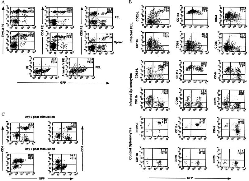
Figure 2.
A subset of activated T cells loses GFP expression after vaccinia infection in vivo or after αCD3 stimulation in vitro. (A) T-GFP mice were infected with vaccinia virus, and on day 7 postinfection, T cells in PELs (Top) and splenocytes (Middle) were tested for GFP expression. PELs also were tested for cell viability (Bottom). Representative results from one infected mouse (of >10 mice analyzed) are shown. (B) Six days postvaccinia infection, PELs and splenocytes were analyzed for expression of GFP versus activation markers by using three-color flow cytometry. Splenocytes from uninfected mice are shown as a control. Representative results from one mouse each (of four analyzed) are shown after gating on the CD8+ subset. (C) Transgenic splenocytes were stimulated in vitro with αCD3, and GFP expression in the CD4+ and CD8+ subset was monitored over time.

