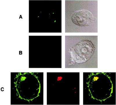
Figure 1.
Fluorophore-conjugated β2m specifically marks internalized class I MHC protein. Untransfected 221 cells incubated with (A) TR-β2m (red) and with FITC-conjugated transferrin (green) or (B) with TR-β2m (red) alone, for 24 hr. (Left) An overlay of red and green fluorescence, and the corresponding Nomarski image (Right). (C) Transfected 221/Cw6-GFP cells incubated with TR-β2m (red) for 2 hr. (Left) Green staining of Cw6-GFP, (Middle) red staining of β2m, and (Right) an overlay of fluorescence colors is displayed. (Right) Surface labeling by TR-β2m is time dependent; compare Fig. 2A. Yellow indicates colocalization of green and red staining. Magnification: ×6,000.

