Figure 2.
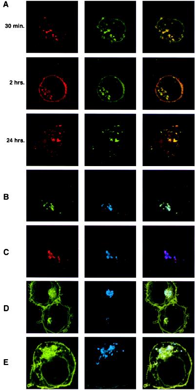
Class I MHC protein cotraffics with class II MHC protein. (A) Intracellular trafficking of exogenously added β2m and superantigen. 221/Cw6 cells incubated with TR-β2m (red) and FITC-conjugated SEA (green) for 30 min, 2 hr, and 24 hr. (B) 221/Cw6 cells incubated with Alexa 488-β2m (green) and Cy5-conjugated SEB (blue) for 2 hr. (C) 221/Cw6 cells incubated with TR-β2m (red) for 2 hr, then fixed, permeabilized, and stained with Cy5-conjugated mAb TÜ39 (blue). (D) 221/Cw6-GFP cells incubated with Cy5-SEB (blue) for 2 hr. HLA-Cw6-GFP (green), Cy5-SEB (blue), and an overlay of both is shown. (E) 221/Cw6-GFP cells fixed, permeabilized, and stained with Cy5-conjugated mAb TÜ39. HLA-Cw6-GFP (green), Cy5-TÜ39 (blue), and an overlay of both is shown. (Right) Yellow indicates colocalization of green and red staining, white or light blue indicates colocalization of green and blue staining, and purple indicates colocalization of red and blue staining. Magnifications: (A–C and E) ×6,000; (D) ×4,000.

