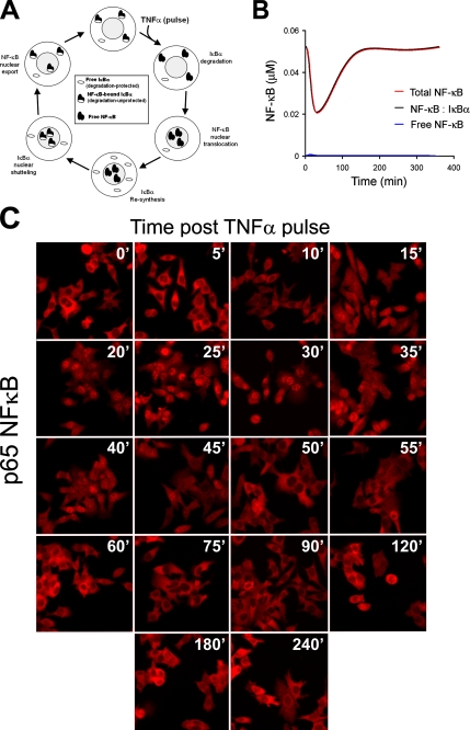
FIGURE 5.
Nuclear export of NF-κB may regulate IκBα sensitivity to ligand-induced degradation. A, a schematic illustrating subcellular localization and levels of free IκBα, free NF-κB, and NF-κB-bound IκBα in response to a pulse of TNFα. B, computationally predicted profile of all cytoplasmic populations of NF-κB following a pulse of TNFα. Note the exceedingly small free NF-κB population. C, HepG2 cells were stimulated with a 30-s TNFα pulse. At the indicated time points, cells were fixed, permeabilized, and immunostained for p65 NF-κB. Shown are representative immunofluorescence confocal photomicrographs.

