Figure 1.
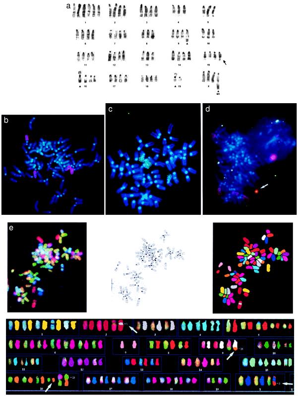
G banding, chromosome painting, FISH, and SKY prove that DCPC21 is a translocation-negative PCT. (a) G banded karyotype of DCPC21 lacking any PCT-associated chromosomal translocations involving chromosomes 12 (IgH), 6 (IgK), 16 (IgL), and 15 (c-myc). The duplicated band on one chromosome 15 (arrow) was mapped to band 15D2, where c-myc is located. Additional chromosomal aberrations (see ▴), such as the elongated chromosome 9 and the marker chromosome M1 as well as the centromerically fused Rb16;19 and the Rb19 isochromosome, probably are acquired during neoplastic progression (see text). (b and c) Chromosome painting of DCPC21 metaphases with chromosome 12 (red) (b) and with chromosome 15 paint (green) (c). No translocation between chromosomes 12 and 15 is visible. In addition, no chromosome 12- or 15-derived material is found as part of any other chromosome. (d) Painting of a DCPC21 metaphase with chromosome 12 (red) and FISH with c-myc (green). The arrow points a large EE that hybridizes with red and green, indicating the presence of chromosome 12-derived sequences and c-myc on the EE. (e) SKY analysis of a DCPC21 metaphase. The data are presented as follows. The image in the upper left corner of the composite shows a representative metaphase obtained with the Spectra Cube before the classification of the spectral colors. The upper center image shows the inverted 4′,6-diamidino-2-phenylindole (DAPI) banding of the same metaphase plate, and the image in the upper right corner displays the spectral colors as classified by skyview 1.2 (Applied Spectral Imaging). The lower image shows identical chromosome pairs of nonclassified and classified DCPC21 chromosomes. SKY corroborates the results of the G banding and chromosome painting: DCPC21 PCT cells do not exhibit any chromosomal translocation. SKY revealed the translocation of chromosome 16-derived material onto the telomeric part of chromosome 9 (see arrows) and the insertion of a chromosome 3-derived band into chromosome 2 (arrow). A centromeric fusion occurred between chromosomes 16 and 19 (Rb 16;19). The M1 marker contains both chromosome X- and 5-derived chromosomal segments (arrow).

