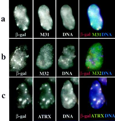
Figure 5.
Localization of β-gal/18D6 fusion, M31, M32, and ATRX proteins in mouse (F9/18D6) nuclei. Immunostaining of F9/18D6 nuclei with antibody against β-gal (images on the far left and red in merged images), together with antibodies against M31 (a), M32 (b), and ATRX (c) (second from left and green in merged images), is shown. DNA was counterstained with DAPI (second from right and blue in merged images). Colocalized signals appear as white.

