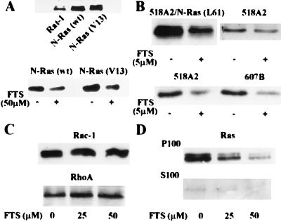
Figure 1.
FTS induces a decrease in the amount of N-Ras in Rat-1 fibroblasts and human melanoma cells without an effect on the amount of Rac-1 and Rho A. Cells were plated at a density of 1.5 × 10⁶ cells per 10-cm dish in DMEM/10% FCS. After 24 h, the cells received either 0.1% DMSO carrier solution (control) or FTS. Ras, Rac-1, and Rho A expression were then determined in total cell lysates (10 μg of protein) by immunoblotting/enhanced chemiluminescence assays with the corresponding antibodies as detailed in Materials and Methods. (A Upper) Immunoblots with pan-Ras Ab demonstrating the amounts of Ras in untransfected Rat-1 cells and Rat-1 cells stably expressing wild-type N-Ras or activated N-Ras (V13). (Lower) Immunoblots with pan-Ras Ab demonstrating the reduced amount of N-Ras in N-Ras expressing Rat-1 cells after a 24-h treatment with 50 μM FTS. (B Upper) Typical immunoblots with pan-Ras Ab demonstrating the FTS-induced reduction of N-Ras in human 518/N-Ras (L61) melanoma cells and the drug-induced reduction in the amount of endogenous Ras in 518A2 cells. (Lower) Immunoblots with N-Ras Ab demonstrating the FTS-induced reduction in the amount of N-Ras in 518A2 and in 607B cells after 24 h of FTS treatment. The results shown are representative examples of three sets of experiments. (C) Immunoblots with Rac-1 and Rho A Abs demonstrating the lack of FTS effects on the total amounts of Rac-1 and Rho A in 518A2 cells after a 24-h treatment with FTS at the indicated concentrations. (D) Dislodgment of Ras by FTS from 518A2 cell membranes. Cells were treated with the indicated concentrations of FTS for 24 h and Ras was then determined in the particulate (P100) and cytosolic (S100) fractions with pan-Ras Ab as detailed in Materials and Methods.

