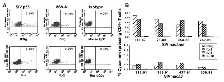
Figure 5.
Intracellular cytokine analysis of CD4+ T cells after stimulation with SIV p55. (A) PBMCs from an SIVmac239Δnef-immunized macaque were stimulated with SIV p55- or VSV-N-pulsed autologous adherent cells for 24 h, and then intracellular expression of IL-2 and IFN-γ in CD4+ T lymphocytes was evaluated by flow cytometry. The numbers shown in the upper right-hand corner of each panel in A correspond to the percentages of CD4+ T cells expressing the given cytokine. (B) Intracellular cytokine expression by SIV p55-specific CD4+ T cells from macaques infected with live attenuated SIV strains.

