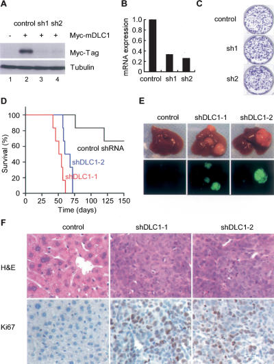
Figure 2.
DLC1 loss triggered by in vivo RNAi promotes tumorigenesis in a mouse liver cancer model. (A) Immunoblots of 293T cells cotransfected with a 6xMyc-tagged murine dlc1 cDNA (lanes 2–4) and a control shRNA (lane 2) or DLC1 shRNAs (lanes 3,4). Tubulin serves as a loading control. (B) DLC1 qRT–PCR of p53-null liver progenitor cells coinfected with Myc and a control shRNA or two DLC1 shRNAs. (C) Cells as in B were plated at equal number and stained with crystal violet after 8 d. Shown are representative results from three experiments. (D) DLC1 loss cooperates with Myc and p53 loss to accelerate liver tumor formation. Kaplan-Meier survival curve of mice transplanted with p53-null liver progenitor cells coinfected with Myc and DLC1 shRNAs (n = 6 for each group). (E) Representative images of explanted livers. GFP imaging identifies shRNA transduced cells. (F) Histopathology of representative tumors. Proliferating cells were labeled by Ki67 staining.

