Figure 1.
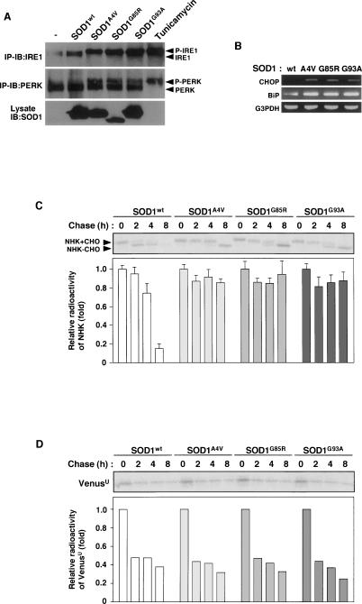
SOD1mut triggers ER stress and inhibition of ERAD. (A) NSC34 cells were lysed after infection with Ad-SOD1wt, Ad-SOD1A4V, Ad-SOD1G85R, or Ad-SOD1G93A for 48 h or treatment with 2.5 μg/mL tunicamycin (a potent inducer of ER stress) for 2 h and analyzed by immunoprecipitation-immunoblotting (IP-IB) with antibodies to IRE1α or PERK. (P-IRE1) Activated IRE1; (P-PERK) activated PERK. The presence of SOD1 in the same lysates is shown. (B) NSC34 cells were transfected with SOD1wt or SOD1mut for 48 h. Expression of BiP and CHOP was examined by RT–PCR. (C) NSC34 cells were transfected with NHK and SOD1wt or SOD1mut. Cells were pulse-labeled with [35S] methionine and cysteine and chased for the indicated time periods. Cell lysates were immunoprecipitated with antibody to α1AT. (NHK + CHO) Glycosylated NHK; (NHK − CHO) deglycosylated NHK. The relative radioactivities in NHK at different times of chase were calculated and are shown as fold decreases relative to the intensity observed at 0 h chase. (D) NSC34 cells were transfected with VenusU and SOD1wt or SOD1mut. Cells were pulse-labeled with [35S] methionine and cysteine and chased for the indicated time periods. Cell lysates were immunoprecipitated with antibody to GFP. The relative radioactivities in VenusU at different times of chase were calculated and are shown as fold decreases relative to the intensity observed at 0 h chase.

