Figure 7.
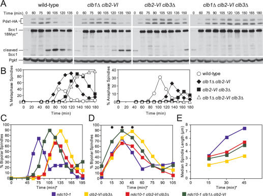
clb2-VI clb3Δ cells are defective in Securin degradation and anaphase spindle elongation. (A,B) Wild-type (A1951), clb1Δ clb2-VI (A15111), clb2-VI clb3Δ (A19136), and clb1Δ clb2-VI clb3Δ (A1938) cells, each carrying Pds1-HA and Scc1-18Myc, were grown and treated as described in Figure 1B. (A) The amounts of full-length Scc1-18Myc (Scc1-18Myc*), the C-terminal cleavage fragment of Scc1-18Myc (cleaved Scc1), and Pds1-HA were also examined. Pgk1 was used as a loading control in Western blots. (B) The percentage of cells with metaphase (left graph) and anaphase (right graph) spindles was determined at the indicated times. (C–E) ndc10-1 (A2733), clb2-VI clb3Δ (A2887), ncd10-1 clb2-VI clb3Δ (A19288), and ncd10-1 clb1Δ clb2-VI (A17126), each carrying a Cdc14-3HA fusion, were grown and treated as described in Figure 1B. The percentage of cells with bipolar spindles is shown in C (n = 200). In the graph shown in D, the time points were adjusted so that onset of bipolar spindle formation occurred concomitantly. The time points chosen for analysis were marked with a black circle. For the ndc10-1 strain, the 0 time point in D corresponds to the 45-min time point in C; for the ncd10-1 clb1Δ clb2-VI strain, the 0 time point corresponds to the 75-min time point in C; for strains clb2-VI clb3Δ and ncd10-1 clb2-VI clb3Δ the 0-min time points correspond to the 90-min time point in C. The delay in bipolar spindle assembly in the ncd10-1 clb1Δ clb2-VI strain relative to ndc10-1 strain is not reproducible and not characteristic of this mutant. (E) The median spindle length of each strain. The time points correspond to the time points in D marked with a black circle. At least 140 cells were examined per time point. Box and whisker plots of spindle lengths from this experiment are shown in Supplemental Figure 3D.

