Abstract
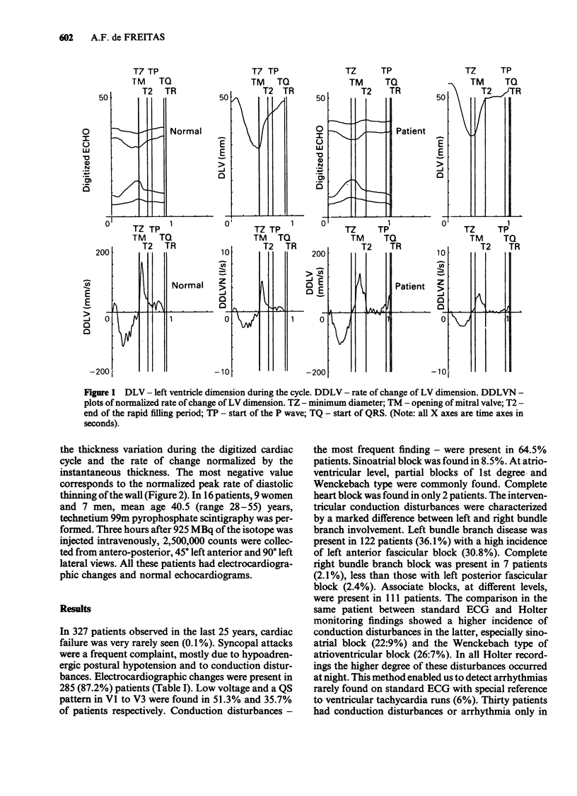
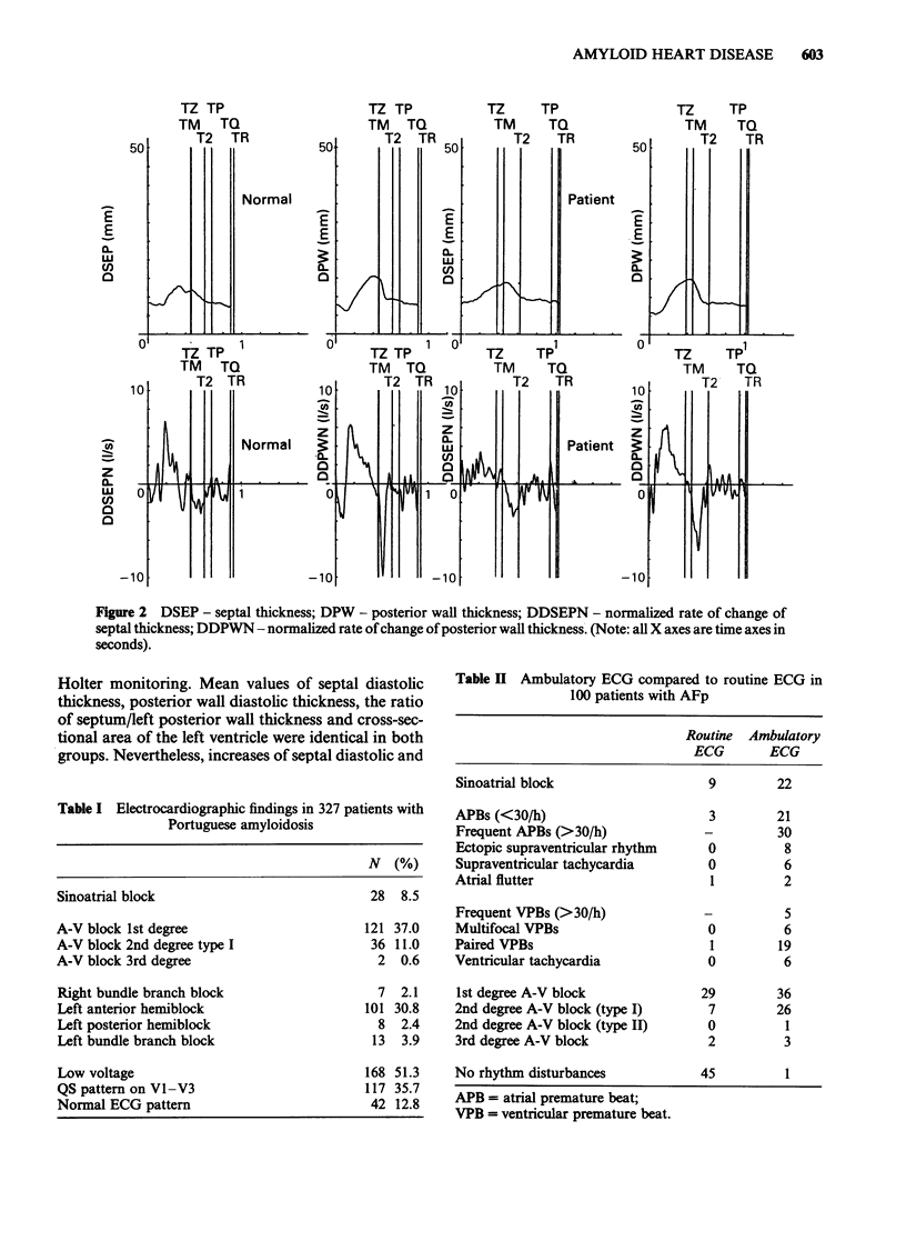
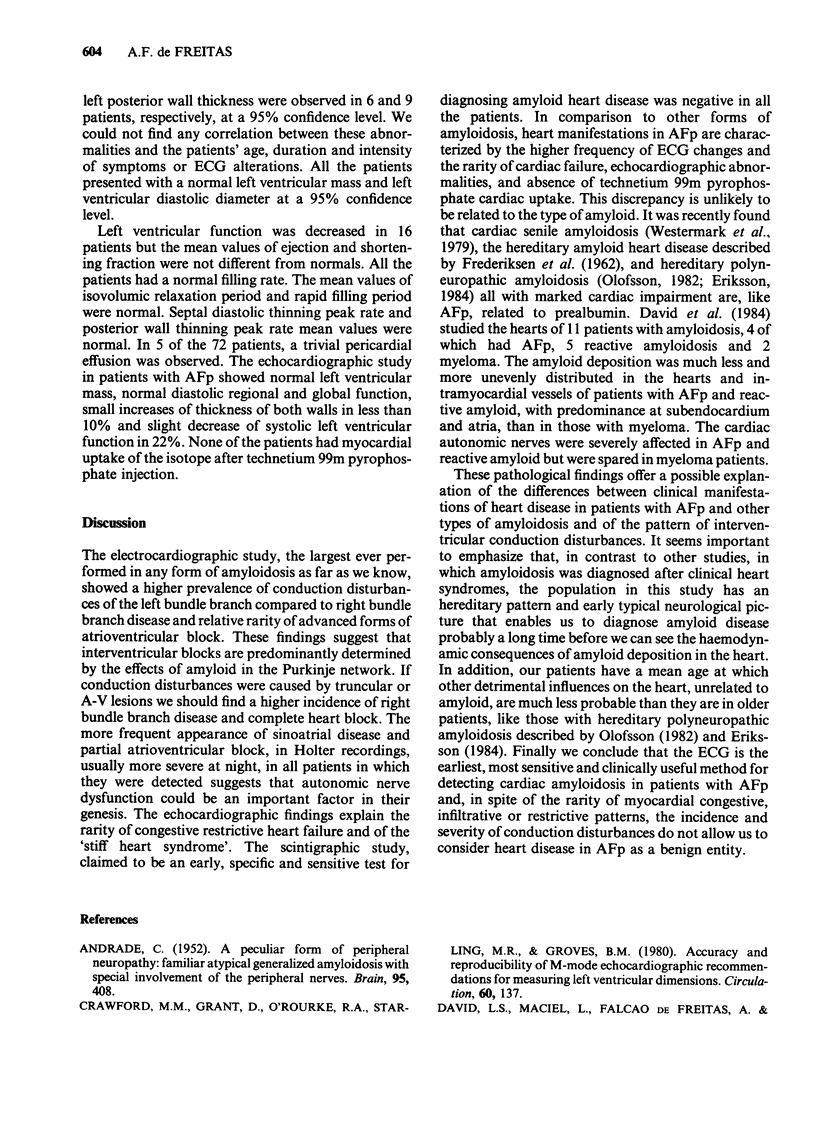
A systematic investigation was performed in patients with familial amyloidotic polyneuropathy, Portuguese type (AFp) to assess the pattern and incidence of cardiac involvement. Of 327 patients investigated, ECG abnormalities were present in 285 (87.2%). Low voltage and QS pattern in V1, V2, V3 were found in 51.3% and 35.7% patients respectively. Conduction disturbances were present in 211 (64.5%). Sinus node disease, 1st degree and Wenckebach interventricular blocks were frequent. Complete atrioventricular block was observed in only 2 patients (0.6%). Left anterior hemi-block was present in 30.8%, left bundle branch block in 3.9%, left posterior hemi-block in 2.4% and right bundle branch block in 2.1%. Holter monitoring showed a much higher incidence of conduction disturbances, most of these occurring at night. The mean values of septum and posterior wall thickness and mass evaluated by echocardiography in 72 patients were normal. The systolic and diastolic global and regional functions, determined in 12 patients, analysing the echo by a digitization computer technique, were normal. In 7% a trivial pericardial effusion was observed. In 16 patients with ECG changes and normal echocardiograms the technetium 99m pyrophosphate scanning was negative. We conclude that the ECG is the most precise, sensitive and clinically useful method for detecting cardiac amyloidosis in patients with AFp. In spite of the rarity of congestive and restrictive patterns, the incidence and severity of conduction disturbances does not allow us to consider heart disease in AFp as a benign entity.
Full text
PDF


Selected References
These references are in PubMed. This may not be the complete list of references from this article.
- ANDRADE C. A peculiar form of peripheral neuropathy; familiar atypical generalized amyloidosis with special involvement of the peripheral nerves. Brain. 1952 Sep;75(3):408–427. doi: 10.1093/brain/75.3.408. [DOI] [PubMed] [Google Scholar]
- Crawford M. H., Grant D., O'Rourke R. A., Starling M. R., Groves B. M. Accuracy and reproducibility of new M-mode echocardiographic recommendations for measuring left ventricular dimensions. Circulation. 1980 Jan;61(1):137–143. doi: 10.1161/01.cir.61.1.137. [DOI] [PubMed] [Google Scholar]
- FREDERIKSEN T., GOTZSCHE H., HARBOE N., KIAER W., MELLEMGAARD K. Familial primary amyloidosis with severe amyloid heart disease. Am J Med. 1962 Sep;33:328–348. doi: 10.1016/0002-9343(62)90230-9. [DOI] [PubMed] [Google Scholar]
- St John Sutton M. G., Reichek N., Kastor J. A., Giuliani E. R. Computerized M-mode echocardiographic analysis of left ventricular dysfunction in cardiac amyloid. Circulation. 1982 Oct;66(4):790–799. doi: 10.1161/01.cir.66.4.790. [DOI] [PubMed] [Google Scholar]
- Westermark P., Johansson B., Natvig J. B. Senile cardiac amyloidosis: evidence of two different amyloid substances in the ageing heart. Scand J Immunol. 1979;10(4):303–308. doi: 10.1111/j.1365-3083.1979.tb01355.x. [DOI] [PubMed] [Google Scholar]


