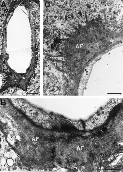
Figure 2.
Ultrastructure of vascular amyloid in APP23 mice. (A) Cortical vessel of a 20-month-old hemizygous APP23 mouse that is surrounded by amyloid (arrowheads). Boxed area is shown in B. (B) High power view of amyloid fibrils (AF) between the endothelial cells (E) and the neuropil. (C) Fine radiating amyloid fibrils often infiltrate the nearby neuropil (arrowheads). Such infiltrating fiber-bundles are surrounded by a dense granular cytoplasm. [Bars = 5 μm (A) and 1 μm (B and C).]

