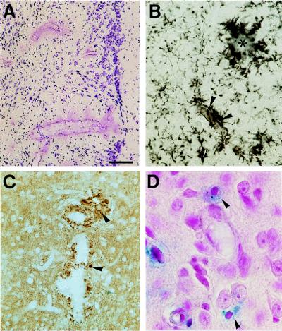
Figure 4.
Neurodegeneration associated with vascular amyloid in APP23 mice. (A) Cresyl violet staining suggests neuron loss surrounding vessels with extensive vascular amyloid. (B) Activated microglia were observed when amyloid was present in the neuropil either in the form of plaques (asterisk) or dyshoric amyloid-containing vessels (arrowheads). (C) Synaptophysin-labeling reveals dystrophic boutons (arrowheads) around vascular amyloid infiltrating the neuropil. (D) Staining for iron (blue) shows microglia that have incorporated products from the blood—an indication of microhemorrhage. [Bars = 40 μm (A), 60 μm (B), 13 μm (C), and 10 μm (D).]

