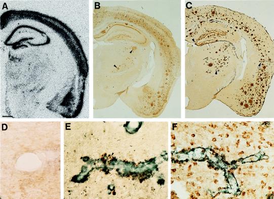
Figure 5.
Regional and neuron-specific expression of human APP in APP23 mice. (A) In situ hybridization for human APP reveals labeling in neocortex, hippocampus, and amygdala. Other regions, such as the thalamus, had no detectable APP expression. (B) Similarly, immunohistochemistry with an antibody specific to human APP reveals neuron labeling in the same regions. However, labeling of dystrophic boutons in the thalamus is also apparent (arrowheads). (C) Immunohistochemistry with an antibody to Aβ reveals vascular amyloid (arrowheads) and amyloid plaques in neocortex, hippocampus, amygdala, and also thalamus. (D) A high magnification view of a thalamic vessel stained for human APP indicates no expression of the transgene within the vasculature. (E and F) Amyloid-laden vessels are shown at high magnification for comparison of human APP expression (brown) and Aβ deposition (blue–gray) between thalamus (E) and neocortex (F). In the cortex, clear localization of APP is apparent within neurons and in dystrophic neurites around amyloid. In the thalamus, in contrast, no neuronal labeling is apparent, although APP is also localized within dystrophic neurites (arrowhead). [Bars = 500 μm (A–C), 10 μm (D), and 30 μm (E and F).]

