Figure 6.
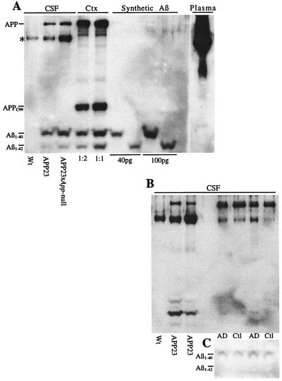
High levels of human Aβ in CSF of APP23 mice. (A) Western blot for human Aβ in CSF (1 μl) from a nontransgenic control [wild-type (Wt)], APP23, and APP23 × App-null mouse, with cortex from an APP23 mouse and synthetic Aβ shown for comparison. The wild-type mice showed no reactivity to human Aβ. In contrast, APP23 and APP23 × App-null showed CSF levels of Aβ1–40 of ≈40 pg/μl. Aβ1–42 was also detectable; however, levels were much lower than Aβ1–40. APP (secreted and full length) was also detectable, and, in CSF, the ratio of Aβ to APP was much higher than in cortex, indicating the presence of cellular APP forms in cortex. When the blot was incubated with a C-terminal antibody (C8; not shown), the APP and Aβ bands from the CSF samples were no longer present, indicating that the APP band in CSF represents secreted APP. A nonspecific band (*) was observed in all CSF samples, which showed no relationship to either APP or Aβ levels and was highly variable from experiment to experiment. A 2-μl plasma sample from an APP23 mouse is shown with no detectable levels of Aβ indicated. (B) Comparison of Aβ and APP levels between mouse and human CSF. Again, high levels of Aβ were present in CSF (2 μl) from the two additional APP23 mice shown, which was not seen in the WT mouse. CSF samples from two AD patients and two aged human controls had much lower Aβ levels than the transgenic mice. Interestingly, the human CSF had a higher ratio of APP to Aβ, perhaps because of the Swedish mutation used in APP23 mice. (C) To better measure Aβ levels in human CSF, five times the volume was loaded (10 μl) and indicated values <5 pg/μl.

