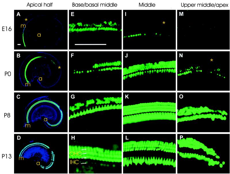
Figure 2.
Developmental pattern of GFP expression in hair cells of cochlea in transgenic mice from E16 to P13. (A–D) Flat-mounted cochlea of the apical half of the cochlea at E16, P0, P8, and P13, respectively. GFP epifluorescence (green) is superimposed on the cochlear outline visualized by DIC (blue). Gaps at P8 and P13 are preparation artifacts. a, apex; m, middle turn. (E–H) Immunodetection of GFP on flat mounts of the base/basal part of the middle turns of the cochlea at E16, P0, P8, and P120 (adult), respectively. (I–L) GFP immunodetection on flat mounts of the middle turns of the cochlea at E16, P0, P8, and P13, respectively. (M–P) Immunodetection of GFP on flat mounts of the upper middle turns at E16, P0, and the apical turns at P8 and P13, respectively. Stars in A and I, B and N correspond to similar positions. A–D are at the same magnification, and E–P are at the same magnification. Bars = 100 μm.

