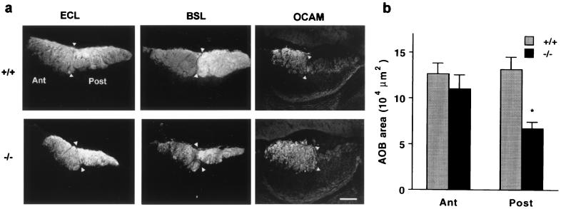
Figure 4.
The posterior AOB fiber layer is reduced in Go −/− mice. (a) Parasagittal sections of the P15 AOB from control and Go knockout mice were stained with the lectins ECL or BSL or with anti-OCAM antibodies. Anterior (Ant), posterior (Post), and the border of the two AOB lobes (arrowheads) are indicated. In the Go −/− sections, the posterior half is reduced in area, but the staining characteristics are similar to those of control AOB. (Bar = 100 μm.) (b) The area of the two halves of the AOB revealed by ECL staining was measured in mid AOB sections from P15 mice. The area of the posterior AOB is significantly decreased in the Go −/− mice (*, P ≤ 0.05, Student’s two-tailed t test). The data are means ± SEM from 5–8 animals in each group.

