Figure 6.
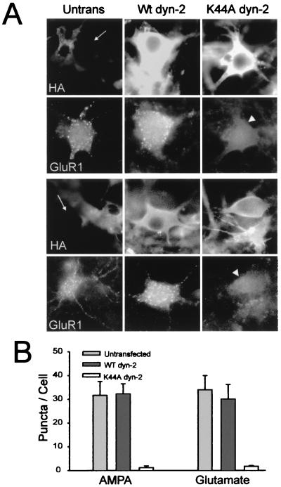
Ligand-induced internalization of AMPARs is dynamin-dependent. HA-tagged wild-type or K44A mutant dynamin−2 were expressed in hippocampal cells via adenovirus-mediated transfection. Neurons were then examined for AMPAR internalization. (A) Micrographs showing the specific inhibition of AMPAR internalization caused by K44A mutant dynamin. The top two rows illustrate cells in which AMPAR internalization was induced by 100 μM AMPA for 15 min, conditions that induce NMDAR-independent internalization of AMPARs. The bottom two rows illustrate the same experiment conducted with the pulse–chase protocol with 10 μM glutamate applied for 1 min, conditions that reveal NMDAR-dependent internalization of AMPARs. In each set of panels, expression of HA-tagged dynamin constructs is indicated (HA), and internalized AMPARs detected in the same cells are shown (GluR1). With either protocol, substantial internalization of AMPARs was observed in cells not expressing mutant dynamin (Left, Untrans; arrow indicates neuron that has no detectable HA-tagged dynamin expression) or in neurons expressing HA-tagged wild-type dynamin−2 (Center, Wt dyn-2). In contrast, in cells expressing K44A mutant dynamin−2 (Right, K44A dyn-2, arrowhead), internalization of AMPARs was strongly inhibited. (B) Quantitation of AMPAR internalization induced by both ligand-activation protocols (n = 8 for AMPA application and n = 15 for glutamate application).

