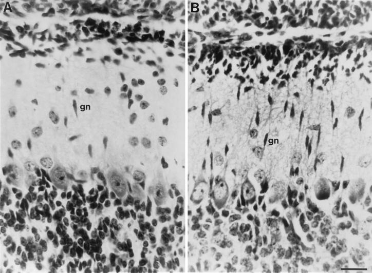
Figure 1.
Granule cells in the molecular layer of P10 cerebella from tPA+/+ and tPA−/− mice. (A) Sagittal section from tPA+/+ mouse showing elongated, presumptive migrating, granule neurons (gn) in the cell sparse molecular layer. (B) Sagittal section of an identical folia and similar lateral displacement as in A, from a tPA−/− mouse showing large numbers of elongated migratory granule neurons in the cerebellar molecular layer. About 2- to 3-fold more granule neurons are seen in the molecular layer of tPA−/− mice, where granule cell migration appears delayed. (Bar = 50 μm.)

