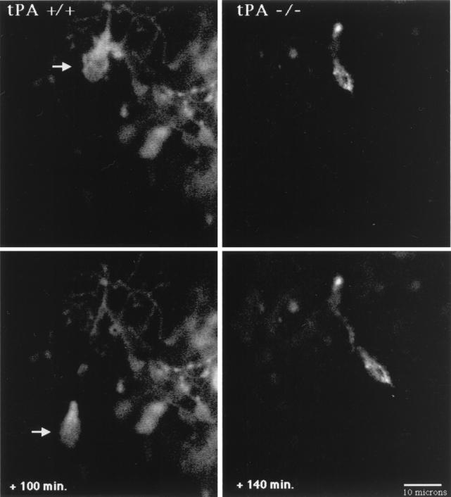
Figure 3.
Confocal microscopy images of migrating granule neurons in cerebellar slices from tPA+/+ and tPA−/− P8 mice. In the tPA+/+ mouse (Left), a DiI-labeled granule neuron (arrow) migrates inward at 15 μm/h from the EGL over the next 100 min, leaving behind its trailing axon. The P8 cerebellar slice from a tPA−/− mouse (Right) shows an inwardly migrating granule neuron that moves through the molecular layer at 5.6 μm/h over the next 140 min. Digital images were collected at 20- to 30-min intervals. (Bar = 10 μm.)

