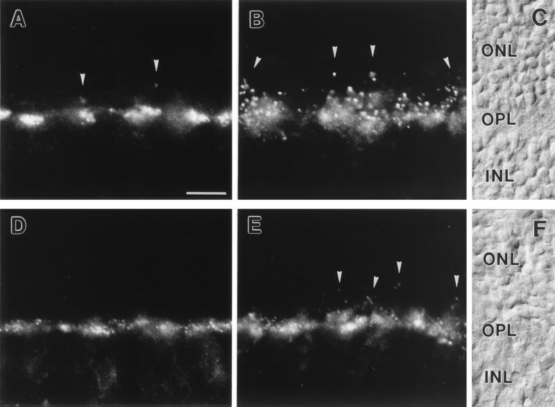
Figure 2.
(A–C) Micrographs of vertical cryostat sections of mouse retina stained with an antiserum against GluR1 (A) and against GluR2 (B). (C) Nomarski micrograph showing the retinal layers (ONL, outer nuclear layer; OPL, outer plexiform layer; INL, inner nuclear layer). (D–F) Micrographs of vertical cryostat sections of rat retina stained with an antiserum against GluR1 (D) and against GluR2 (E). (F) Nomarski micrograph showing the retinal layers. For both species and for both glutamate receptor subunits, strong immunofluorescence was found in the OPL. The labeling was mainly associated with cone pedicles (the large clusters) but was also found at rod spherules (arrowheads). Bar = 10 μm in A for A–F.

