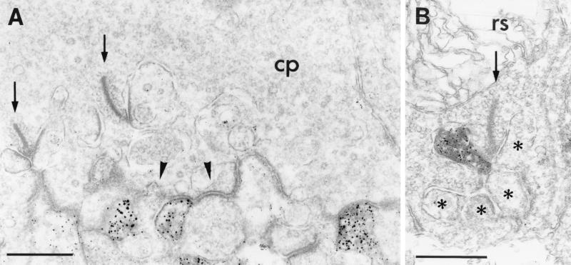
Figure 3.
(A) Electron micrograph showing the postsynaptic localization of GluR1 at a cone pedicle (cp) in the OPL of mouse retina. GluR1 immunoreactivity was found on some dendrites of OFF-cone bipolar cells making flat noninvaginating synaptic contacts at the cone pedicle; other flat contacts were not labeled (arrowhead). (B) Electron micrograph showing the localization of GluR2 on a horizontal cell process postsynaptic at a rod spherule (rs) in the OPL of rat retina. The presynaptic ribbons in the cone pedicle and in the rod spherule are marked by arrows; the unlabeled postsynaptic elements at the rod spherule are marked by asterisks. Bar = 0.5 μm in A and B.

