Figure 4.
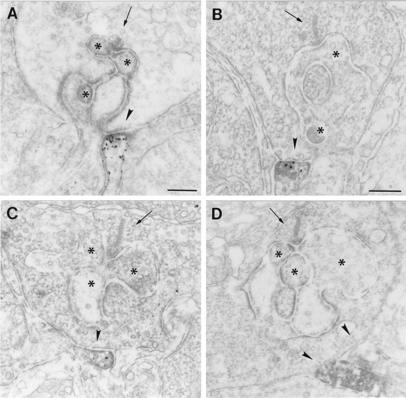
(A–D) Electron micrographs showing flat noninvaginating synaptic contacts (arrowhead) made by putative OFF-cone bipolar cell dendrites at rod spherules in the OPL of mouse (A and C) and rat (B and D) retina labeled for GluR1 (A and B) and for GluR2 (C and D). The presynaptic ribbons in the rod spherules are marked by an arrow. The unstained horizontal cell processes and dendrites of rod bipolar cells postsynaptic at the rod synapses are marked by asterisks. Bar = 0.2 μm in A for A, C, and D, and 0.2 μm in B.

