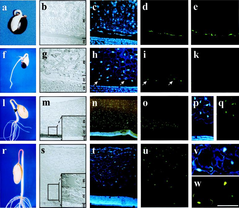
Figure 1.
Nuclear DNA fragmentation in germinating Ricinus endosperm. Sections were prepared from endosperm of 2-day-old (a–e), 3-day-old (f–k), 4-day-old (l–q), and 5-day-old (r–w) seedlings and visualized by using DIC optics (b, g, m, and s; co, cotyledons; cc, collapsed cells; es, living endosperm). Nuclei were stained with DAPI (c, h, n, p, t, and v), and DNA fragmentation was detected by TUNEL (d, i, o, q, u, and w). (e and k) Negative controls of the TUNEL assay in sections from 2-day-old (e) and 3-day-old (k) endosperm. (a–e) Two-day-old seedlings (b) seen by using DIC optics, (c) stained with DAPI, and (d) showing TUNEL-positive cells close to the cotyledons; (e) negative control for TUNEL assay also shows labeling close to the cotyledon. (f–k) Three-day-old seedling (g) seen by using DIC optics, (h) stained with DAPI, and (i) showing TUNEL-positive cells in the first two cell layers next to the cotyledons; (k) negative control for TUNEL assay shows no labeling on endosperm cells. (l–q) Four-day-old seedling (m) seen by using DIC optics showing the whole endosperm with attached cotyledons, (n) stained with DAPI, (o) showing TUNEL-positive nuclei with no labeling on the cotyledons, (p) higher magnification of a section stained with DAPI, and (q) same section as p showing TUNEL-positive cells exclusively in the endosperm. (r–w) Five-day-old seedling (s) section of whole endosperm with attached cotyledons, (t) stained with DAPI, (u) showing TUNEL-positive cells in the whole endosperm with no labeling on the cotyledons, (v) higher magnification of nuclei stained with DAPI, and (w) same section as v shows TUNEL-positive cells. [Scale bar: (a and f) 2 cm; (l and r) 3.5 cm; (m–o) 800 μm; (s–u) 400 μm; (b–e, h, i, k, p, and q) 200 μm; (g, v, and w) 75 μm.]

