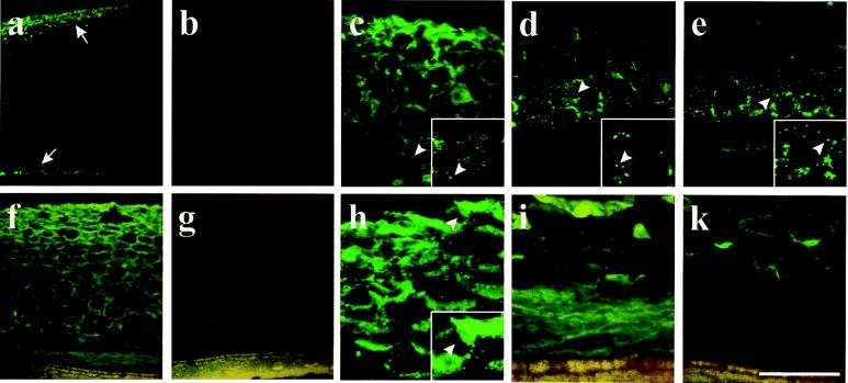
Figure 3.
Development of ricinosomes and cellular distribution of cysteine endopeptidase in germinating Ricinus endosperm. Sections from 3-day-old (a–e) and 5-day-old (f–k) germinating endosperm were labeled with α-Cys-EP antibody (a, c, d, f, h, and i), the preimmune serum (b and g), and the α-propeptide antibody (e and k) by using a FITC-labeled secondary antibody. (a) Cys-EP is detected (arrows) in the outer part of the endosperm and close to the cotyledons in 3-day-old seedlings. (b) Preimmune serum. Higher magnification of the outer part (c) and the inner part (d) of the endosperm shows a punctate labeling in ricinosomes. (e) The α-propeptide antibody labels ricinosomes in the inner part of the endosperm in a punctate pattern. (f) Detection of Cys-EP in the endosperm of 5-day-old seedlings. (g) Preimmune serum. (h) Higher magnification of the outer part of the endosperm shows a mostly diffuse labeling with only a few ricinosomes. (i) Higher magnification of the inner part of the endosperm shows diffuse labeling over the collapsed cells. (k) No labeling is observed over the collapsed cells with the α-propeptide antibody. Arrows in a indicate labeling in the outer and the inner part of 3-day-old endosperm. Arrowheads in c–e and h indicate single ricinosomes. (Insets, c–e and h) A further 2-fold magnification of the region around the arrowheads. [Scale bar: (a and b) 800 μm; (f and g) 400 μm; (k) 200 μm; (c–e, h, and i) 100 μm.]

