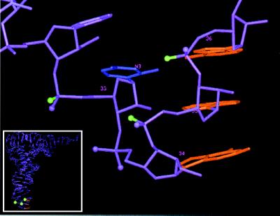
Figure 5.
Three-dimensional structure of the anticodon loop of tRNAPhe from yeast. The three bases of the anticodon are in red. The green balls depict Rp-phosphate oxygens identified in this study which when substituted with sulfur interfere with tRNA P-site binding; those in magenta do not interfere with binding [except at G34 (Rp), at which binding was enhanced]. The base presented in blue is U33; the labeled atom N3 is proposed to interact with the Rp-phosphate oxygen of A36 (32). The complete tRNA is seen in the Inset.

