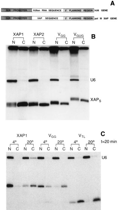
Figure 4.
Expression of XAPs in vivo. (A) Construction of the pol III XAP genes. Restriction sites were introduced at the 3′ end of the hU6 promoter and at the 5′ end of the 3′ flanking region of the hU6snRNA gene. The first 6 bp of the XAP-stem sequence were changed, with respect to the sequence shown in Fig. 2, to facilitate the incorporation of a restriction site sequence into the XAP stem. In addition, the start site was changed to GAG because a higher level of expression was obtained, compared with clones starting with GGG. (B) Intracellular localization of XAPs transcribed by RNA pol III. Equal amounts of hU6 genes and pol III XAP-genes (XAP1, XAP2, VGG, VGUG) were injected into the nucleus of oocytes together with [α-32P]GTP as described (17). Twelve hours later oocytes were separated into nuclear (N) and cytoplasmic (C) fractions, and extracted RNA was analyzed on a denaturing acrylamide gel. (C) Temperature sensitivity of XAP export. XAP1, VGG, and VTL were transcribed in vitro (all uncapped) and injected into the nucleus of oocytes together with U6snRNA. Oocytes were kept for 20 min at 4° or at 20° and separated into nuclear (N) and cytoplasmic (C) fractions, and extracted RNA was analyzed on a denaturing acrylamide gel.

