Figure 5.
LPS Promotes Shedding of GHBP from Adenovirally Expressed GHR in C3H/HeJ and Athymic Nude Mice
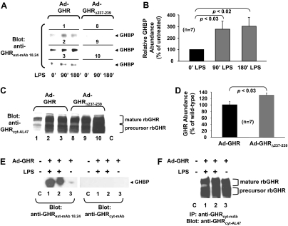
A and B, GHBP shedding. Mice (n = 7 per group) were infected with either Ad-GHR or Ad-GHRΔ237–239, as indicated, both of which are rabbit GHRs. As detailed in Materials and Methods, mice were treated with LPS, and serum obtained from each mouse was precleared with protein G-Sepharose and blotted with a monoclonal antibody that recognizes the rabbit, but not mouse, GHR ECD. Panel A, Blots from three representative mice from each group are shown; panel B, data from the entire Ad-GHR group were densitometrically analyzed and are displayed as mean ± sem. Panels C and D, GHR abundance. Livers from mice analyzed in panels A and B were homogenized, and equal aliquots of cytoplasmic extract were resolved by SDS-PAGE and immunoblotted for GHR. Panel C, Samples from the same three mice per group as were evaluated in A are displayed. The positions of mature and precursor GHR are noted. C is liver cytoplasmic extract from a mouse not infected with either Ad-GHR or Ad-GHRΔ237–239. (Note that at this exposure, endogenous GHR is not detected.) Panel D, Densitometric analysis of the blots in panel C. Data are displayed as mean ± sem (n = 7) relative to the GHR level in Ad-GHR-infected mice. Panels E and F, GHBP shedding and GHR abundance in nude mice. Panel E, As detailed in Materials and Methods, nude mice were treated as in panel A to detect shed GHBP after infection with Ad-GHR or control, as indicated. Serum samples were blotted with anti-GHRext-mAb18.24 and, as a negative control, anti-GHRcyt-mAb. Note LPS-induced appearance of specifically detected GHBP in serum. Panel F, Livers from the same mice in panel E were extracted, as in panel C, and immunoprecipitated with anti-GHRcyt-mAb. Precipitated proteins were resolved by SDS-PAGE and immunoblotted for GHR. Note similar expression levels of adenovirally expressed GHR and its absence in the control mouse liver.

