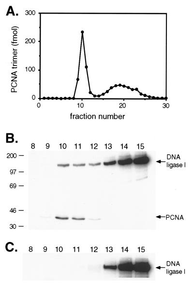
Figure 2.
Formation of DNA–protein complexes by PCNA and DNA ligase I. Nicked plasmid DNA, DNA bound-PCNA, and DNA ligase I were incubated and then fractionated by gel filtration as described in Materials and Methods. (A) Radiolabeled PCNA trimers (2 pmol trimer) that had been loaded onto nicked, circular DNA duplexes (1 μg). 32P-labeled PCNA was detected by liquid scintillation counting. (B) Radiolabeled PCNA trimers (2 pmol trimer) bound to DNA incubated with 32P-labeled DNA ligase I (44 pmol). (C) 32P-labeled DNA ligase I (44 pmol) incubated with nicked, circular DNA duplexes (1 μg). After separation by denaturing gel electrophoresis, labeled proteins were detected and quantitated by PhosphorImager analysis. The positions of DNA ligase I and PCNA are indicated on the right.

