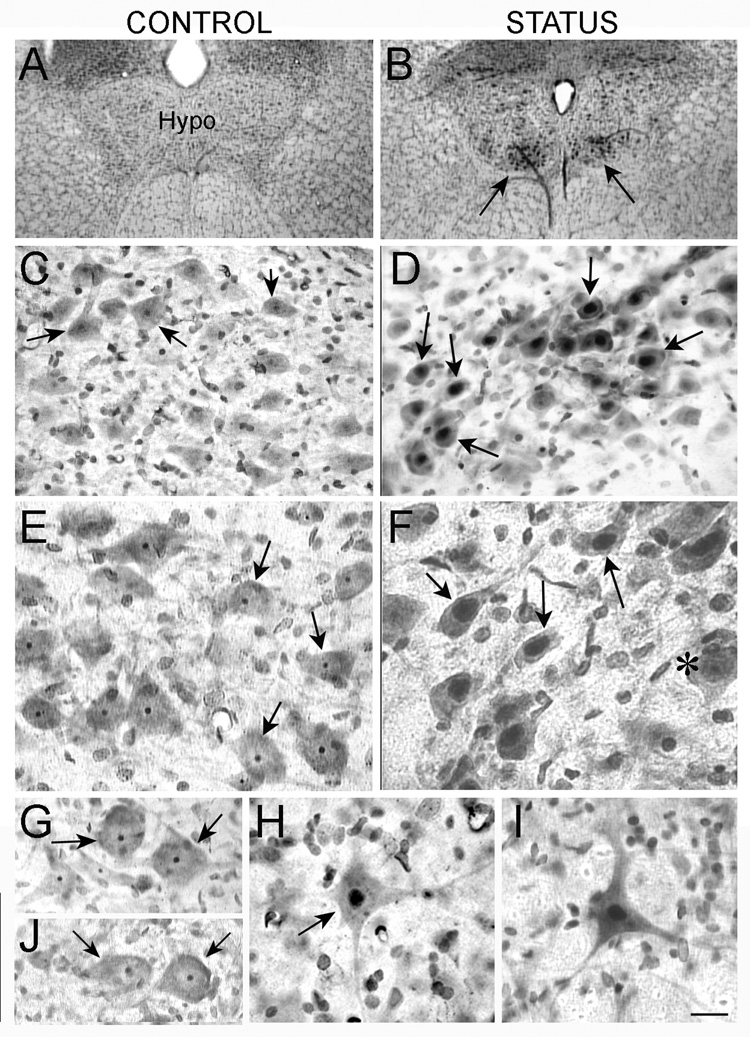
Figure 2. Silver uptake in motor neurons after status.
A–B. Silver stained sections from a saline control (A) and animal that was perfused 1 day after status (B) show increased staining in the hypoglossal nucleus of the animal that had status (arrows in B).
C–D. At higher power, the silver staining in the saline control in A appears to be confined to the nucleolus (arrow in C) or is not evident, but it is in the nucleus after status (arrows in D)
E–F. High magnification and increased contrast is used to illustrate the difference in silver uptake shown in C–D more clearly. In F, an exceptional neuron that does not show silver uptake is marked.
G–H. Silver uptake in spinal cord motor neurons was similar qualitatively to the pattern of staining in hypoglossal motor neurons: the nucleolus was stained in motor neurons from saline-treated controls (arrows in G), and the entire nucleus was stained in motor neurons from animals that had status (arrow in H).
I. A neuron in the facial nucleus with silver stain throughout the nucleolus shows the same distribution of silver as other motor neurons from animals that had status the previous day.
J. Silver uptake in spinal motor neurons 1 week after status epilepticus shows a pattern of silver uptake similar to control rats. Calibration: A–B, 320 µm; C–D, 80 µm; E–J, 40 µm.

