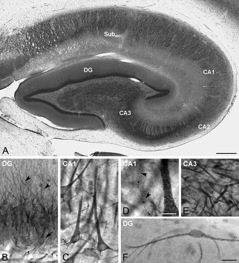
Fig. 1.
Aromatase DAB immunoreactivity in the rhesus monkey hippocampus. (A) Panoramic view of aromatase distribution in the hippocampus (subject 29357). Sub, Subiculum; CA1-CA3 cornu Ammonis subfields 1–3; DG, Dentate gyrus. (B) Aromatase expression in the DG. The image shows aromatase immunostaining both in the perikaryon of some granule cells (arrow) as well as in dendrites that reach the molecular layer (arrowheads) (subject 27697). (C) Aromatase expression in CA1. The image shows several aromatase immunoreactive pyramidal cells (subject 29357). (D) Detail a high magnification of panel C showing an aromatase-immunoreactive fiber (arrowheads). (E) Aromatase expression in CA3. The image shows several neurons expressing aromatase (subject 30691). (F) Aromatase-immunoreactive neuron located in the molecular layer of the dentate gyrus (subject 28816). Scale bars in A: 500 μm; D: 10 μm; F: 25 μm (for B, C, E and F).

