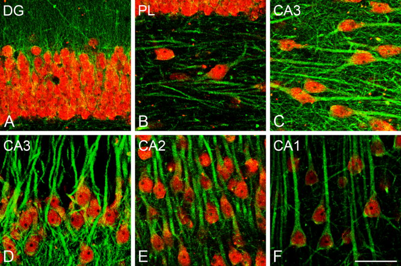
Fig. 2.
Confocal laser scanning microscope (CLSM) images demonstrating colocalization of aromatase (green) and NeuN (red) in the rhesus monkey hippocampus (subject 28816). (A) Colocalization of aromatase and NeuN in the granular cell layer of the DG. (B) Colocalization of aromatase and NeuN in the polymorphic layer (PL) of DG (C,D) Colocalization of aromatase and NeuN in CA3. (E) Colocalization of aromatase and NeuN in CA2. (F) Colocalization of aromatase and NeuN in CA1. Scale bar in F: 50 μm.

