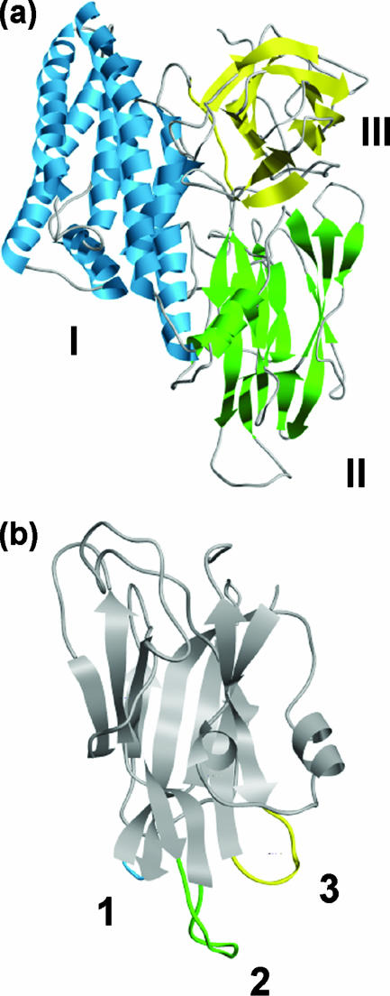
FIG. 1.
The structure of Cry1Aa (30) (PDB code 1CIY) highlighting the three domains of the active toxin (a) and the three apical loops of domain II (b). The molecular graphic images were produced using the UCSF Chimera package (60) from the Resource for Biocomputing, Visualization, and Informatics at the University of California, San Francisco (supported by NIH P41 RR-01081).

