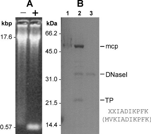
FIG. 5.
TP detection. (A) Phage asccφ28 DNA was restriction digested with ScaI (each ITR has a ScaI site close to its inner end), resulting in two 573-bp terminal DNA fragments. Digested DNA was treated (lane +) or not treated (lane −) with proteinase K and then electrophoresed on a 1.2% agarose gel. (B) Phage asccφ28 DNA (not treated with proteinase K) was electrophoresed on a 12% polyacrylamide gel. Lane 1, asccφ28 DNA carrying the bound TP; lane 2, asccφ28 DNA digested with 1 U of DNase I, which released the TP; lane 3, 1 U of DNase I (control). Lanes 1 and 2 also show that there was residual MCP in the DNA preparation. The N-terminal sequence of the TP was determined and is shown along with the first 10 amino acids of the protein predicted from ORF6 (in parentheses).

