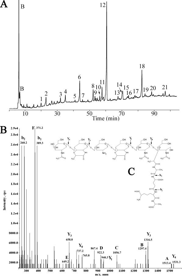
FIG. 3.
HPLC separation and MS analysis of C. perfringens SM101 spore muropeptides. (A) Spore PG was purified, digested with mutanolysin, reduced, and separated using a methanol gradient as previously described (29). Muropeptides were detected by absorbance at 206 nm and are numbered as in Table 4. Peaks labeled B are buffer components. (B) Fragmentation spectrum of muropeptide 18. Ion masses indicated by letters are those predicted in panel C and in Table 5. (C) Structure proposed for muropeptide 18. The arrows indicate fragmentation points to produce the masses indicated in panel B.

