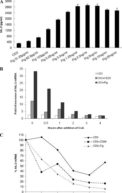
FIG. 4.
IL-2 secretion, mRNA expression, and stability upon costimulation with recombinant B. pseudomallei flagellin. (A) A total of 0.2 × 10⁶ Jurkat cells were stimulated on 2.5-μg/ml anti-CD3 antibody-coated plates alone (CD3) or with the indicated concentrations of B. pseudomallei Flg. A total of 2.0 × 10⁶ Jurkat cells were incubated in the absence of stimulus (untreated), in the presence of 2.5 μg of anti-CD3 antibody/ml alone (CD3), in the presence of anti-CD3 antibody with 1 μg of anti-CD28 antibody/ml (CD3+CD28), or with 1 ng of flagellin/ml (CD3+Flg). After 5 h of stimulation, 0.5 μg of Cs/ml was added to inhibit IL-2 transcription. Total RNA was isolated immediately from cells after 5 h of stimulation (0 h) or 0.5, 1, 2, 3, or 4 h after the addition of Cs. (B) IL-2 mRNA levels in treated samples (CD3, CD3+CD28, and CD3+Flg) at different time points are expressed as the fold increase relative to that of the untreated samples. (C) Within each treatment, the IL-2 mRNA levels in the samples at 0 h are normalized to be 100%; the IL-2 mRNA levels in the samples at 0.5, 1, 2, 3, and 4 h are expressed relative to that at 0 h. The results from one representative experiment out of three are shown.

