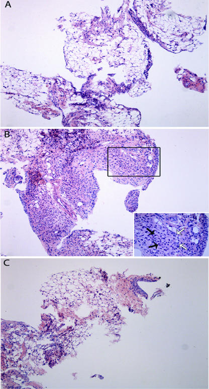
FIG. 4.
Representative illustration of the synovial histopathology of mice subjected to ZYA and treated with the A. suum extract. Mice received 1 mg of A. suum extract (C) or saline (B) given i.p. 30 min prior to 0.1 mg zymosan i.a. Naive mice (A) received saline i.a. All animals were sacrificed at 7 days. The synovia of animals that received just the zymosan display intense and diffuse mononuclear cell infiltration (B) that is clearly reduced in the mice that received the A. suum extract prior to Zy (C). The inset in panel B represents a closer view showing vascular neoformation (black arrows) and macrophage infiltration (white arrows). HE staining. Original magnification, ×100.

