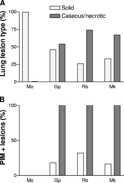
FIG. 2.
Summary of pimonidazole staining by species and lesion type. (A) Summary of the distribution of granulomas, showing solid and caseous histology in each animal model. (B) Proportion of each granuloma type labeling with anti-PIMO in the animal models. Mo, mouse; Gp, guinea pig; Rb, rabbit; Mk, macaque. Open bars, solid granulomas; filled bars, caseous/necrotic granulomas.

