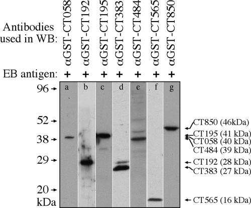
FIG. 3.
Detection of EB proteins with seven anti-fusion protein antibodies. The purified EBs were resolved in SDS-polyacrylamide gel, and the protein bands were transferred onto nitrocellulose membrane for measuring antibody reactivity in a Western blot (WB) assay. Seven mouse anti-GST fusion protein antibodies as listed on top of the figure were used to react with strips of the nitrocellulose membrane (a to g). The molecular masses in kDa are marked along the left, while the antibody-recognized EB protein bands are listed along the right side of the figure. The sizes of the EB proteins are also indicated, in parentheses following each protein's name. Clearly, the seven anti-fusion protein antibodies recognized the corresponding endogenous proteins from the purified EBs. α, anti.

