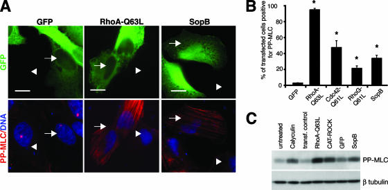
FIG. 10.
SopB expression is sufficient to induce myosin II phosphorylation and is required for activation of myosin II on Sifs. (A) PP-MLC (red) staining in HeLa cells expressing GFP, RhoA-Q63L, or SopB (green). Nuclei are shown in blue. Transfected cells are indicated by arrows, and nontransfected cells are indicated by arrowheads. (B) Percentage of transfected cells positive for PP-MLC staining. Averages ± SD are shown. *, significantly different from GFP-expressing controls (P < 0.05), as determined by one-way ANOVA and Dunnett post hoc analyses. (C) Western blot showing MLC phosphorylation in cells treated with calyculin A, expressing RhoA-Q63L, CAT-ROCK, GFP, or SopB, or mock transfected without DNA (transf. control).

