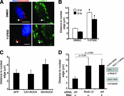
FIG. 5.
Inhibition of ROCK affects SCV positioning. (A) HeLa cells were infected with serovar Typhimurium for 10 min, and then DMSO or the ROCK inhibitor Y-27632 was added to the medium until 2 h p.i. For late-infection experiments, cells were infected for 2 h, and then DMSO or Y-27632 was added to the medium for an additional 6 h. Cells were fixed and immunostained for Salmonella and LAMP-1 and analyzed by immunofluorescence microscopy. Representative images of cells treated with DMSO or Y-27632 are shown. Arrows indicate LAMP-1-positive bacteria. Bars, 10 μm. (B) Samples shown in panel A were analyzed by measuring the distance from the LAMP-1+ bacteria to the closest nuclear edge. Averages ± SE are shown. *, significantly different from the respective DMSO-treated controls (P < 0.05), as determined by a two-tailed, unpaired t test. (C) Cells were transfected with pegfp, pCAT-ROCK, or pDN-ROCK and infected with WT serovar Typhimurium for 8 h. Cells were fixed and immunostained for Salmonella and LAMP-1, and the cells were stained with DAPI as well. The distances from LAMP-1+ bacteria to the closest nuclear edge were determined. Averages ± SD are shown. Asterisks indicate values that are significantly different (P < 0.05) from those of control GFP-transfected cells, as determined by one-way ANOVA and Dunnett post hoc analysis. (D) Cells treated with either control siRNA (ctrl) or siRNA directed against ROCK I and II (Rock I,II) were infected with WT serovar Typhimurium for 8 h. Blebbistatin (Bleb) was also added at 2 h p.i. as indicated. Cells were fixed and immunostained as described above. The distances from LAMP-1+ bacteria to the closest nuclear edge were determined. Averages ± SD are shown. A Western blot demonstrating knockdown of ROCK II in HeLa cells that received ROCK I and II siRNA is shown.

