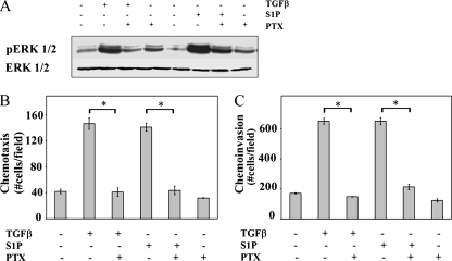
FIG. 4.
ERK1/2 activation, chemotactic migration, and chemotactic invasion are Gi dependent (A to C). OE33 cells were pretreated for 2 h with 100 ng/ml pertussis toxin as indicated. (A) Cells were treated with TGFβ (80 pM) or S1P (50 nM) for 15 min. Cell lysates were separated by SDS-PAGE and analyzed for pERK1/2 and total ERK1/2 by Western analysis. Chemotactic migration (B) and invasion (C) assays were performed as described in Materials and Methods. Data are means ± standard errors of the means from triplicate cultures. Asterisk, P < 0.05.

