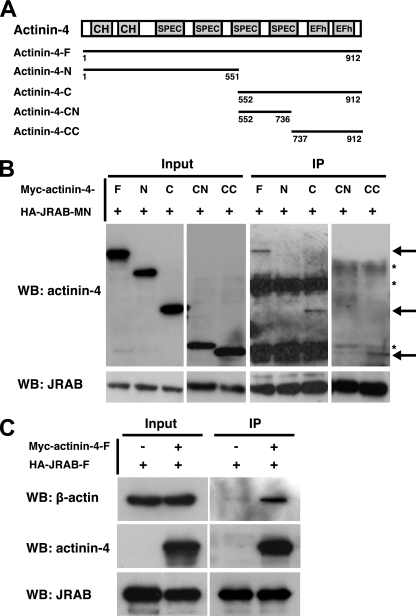
FIG. 3.
Interaction of actinin-4 with JRAB/MICAL-L2 and F-actin. (A) Structures of the full-length and various fragments of actinin-4. Numbers represent amino acid positions. CH, calponin homology domain; SPEC, spectrin repeats; EFh, EF hands. (B) BHK cells cotransfected with pCI-neo-HA-JRAB/MICAL-L2-MN and pCI-neo-Myc-actinin-4-F, pCI-neo-Myc-actinin-4-N, pCI-neo-Myc-actinin-4-C, pCI-neo-Myc-actinin-4-CN, or pCI-neo-Myc-actinin-4-CC were immunoprecipitated (IP) with anti-HA antibody and subjected to Western blotting (WB) analysis using anti-Myc and anti-HA antibodies. Arrows indicate Myc-actinin-4-F, Myc-actinin-4-C, and Myc-actinin-4-CC, and asterisks indicate nonspecific bands. (C) BHK cells cotransfected with pCI-neo-HA-JRAB/MICAL-L2-F and pCI-neo-Myc-actinin-4-F were immunoprecipitated with anti-HA antibody and subjected to Western blotting analysis using anti-β-actin, anti-Myc, and anti-HA antibodies. The results shown in panels B and C are representative of three independent experiments.

