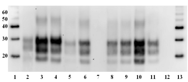
Figure 4.

Glycosylation patterns of PrPres. Western immunoblot of brain samples after treatment with Proteinase-K and using antibody L42 (R-biopharm, diluted 1/2000) to label proteinase resistant PrP. Note significantly (p < 0.001) greater migration of di-, mono- and unglycosylated bands in all 6 clinically affected deer (lanes 5, 6, 8–11) compared to sheep scrapie, experimental ovine BSE and cattle BSE (lanes 2–4 respectively). Also, lack of labelling of protease resistant PrP in negative control deer (lanes 7 and 12). Lane 1 – molecular weight markers (kDa), lane 2 – sheep scrapie, lane 3 – experimental ovine BSE, lane 4 – BSE (inoculum), lane 5 – clinically affected deer 1, lane 6 – clinically affected deer 2, lane 7 – negative control (deer 7 in table 1), lane 8 – clinically affected deer 3, lane 9 – clinically affected deer 4, lane 10 – clinically affected deer 5, lane 11 – clinically affected deer 6, lane 12 – negative control (deer 8 in table 1), lane 13 – molecular weight markers (kDa).
