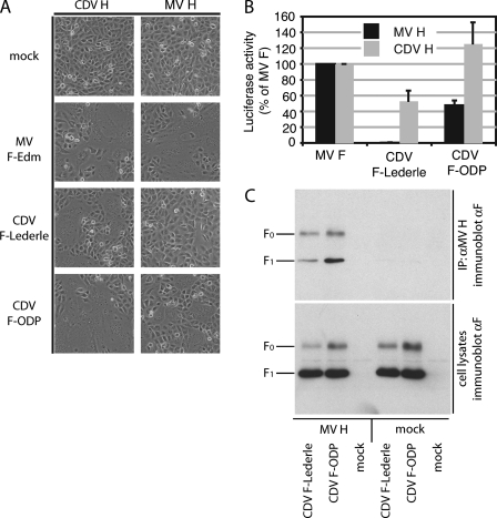
FIGURE 1.
CDV F-ODP but not CDV F-Lederle is triggered by MV H. A, microphotographs of Vero-dogSLAM cells co-transfected with equal amounts of plasmid DNA encoding MV or CDV glycoproteins as specified. The cells were photographed at a magnification of 200× after incubation at 37 °C for seven to 11 h. Mock infected cells expressed only the H protein. B, quantification of cell-to-cell fusion activity using a firefly luciferase reporter-based fusion assay. The values reflect the average luciferase activities of at least three independent experiments ± S.D. per glycoprotein combination and are expressed as the percentages of activity measured for MV F and the respective H. C, CDV F glycoprotein variants show different strengths of interaction with MV H. Co-immunoprecipitation of CDVF-ODP and Lederle with MV H. The lysates of co-transfected cells were subjected to immunoprecipitation using specific antibodies directed against an epitope in the MV H ectodomain. Co-precipitated F (upper panel) was detected in comparison with F present in the lysates prior to precipitation (lower panel) by immunoblotting using a specific antiserum directed against an epitope in the cytosolic tail of CDV F.

